Learning Objectives
By the end of this section, you should be able to:
• Describe the key features and classification of leukemia
• Distinguish between acute and chronic leukemias
• Describe how leukemia affects blood cell function
• Identify key features of leukemias in blood smears
As you work through this section, focus on how changes in leukocyte number, type, and development affect blood cell function and appearance.
Tip: As you read through each passage, click on the underlined terms to see definitions and hear how they are pronounced. This will help reinforce key vocabulary as you work through the material.
Introduction
Leukemia is an uncontrolled proliferation of one type of white blood cell (leukocyte). Like all cancers, leukemic cells typically arise from a single cell that has lost normal control of the cell cycle. As a result, these cells divide uncontrollably and accumulate over time.
Leukemias often involve elevated white blood cell counts, changes in cell maturity, and disruption of normal blood cell production, all of which affect how blood appears and functions.
There are several types of leukemia, reflecting both the different types of white blood cells (five main types) and the stages of development they normally pass through (Kimball, LibreTexts). Leukemia may involve cells from either the myeloid lineage (myelocytic leukemia) or the lymphoid lineage (lymphocytic leukemia). In all cases, the body produces large numbers of abnormal white blood cells that do not function properly (OpenStax, Anatomy and Physiology 2e, Section 18.4).
Leukemias are also classified based on how quickly they progress. In chronic leukemia, more mature leukocytes accumulate and fail to die as they should. In acute leukemia, there is an overproduction of immature, developing leukocytes. In both cases, the cells are typically dysfunctional.
Excessive leukocyte proliferation is known as leukocytosis. Although leukocyte counts are elevated, the cells often lack normal function, leaving the individual more susceptible to infection (OpenStax, Anatomy and Physiology 2e, Section 18.4). In addition, these abnormal cells crowd the bone marrow and bloodstream, interfering with the production of healthy blood cells such as erythrocytes and platelets. This can lead to symptoms such as fatigue, increased risk of infection, and easy bruising or bleeding.
There are several major types of leukemia, including Acute Lymphocytic Leukemia (ALL) and Chronic Myelogenous Leukemia (CML), which we will explore in more detail next. Understanding the differences between these types—particularly CML and AML—is important for recognizing how leukemias present in blood smears and how they affect normal blood cell function.
This section provides background information needed to analyze leukemia case studies.
Acute Lymphocytic Leukemia (ALL)
Acute lymphocytic leukemia is a rapidly progressing cancer that affects lymphocytes, a type of white blood cell involved in the immune response.
In ALL, immature lymphocytes multiply quickly in the bone marrow and bloodstream. Because these cells are not fully developed, they cannot properly fight infections.
ALL is the most common type of leukemia in children, although it can also occur in adults. With modern treatments such as chemotherapy, survival rates for many patients with ALL have improved significantly.
Chronic Myelogenous Leukemia (CML)
One of the most common leukemias is chronic myelogenous leukemia, which is a disease in which the bone marrow makes too many immature white blood cells. CML develops relatively slowly compared to acute leukemias. It begins in a bone marrow stem cell and primarily affects the myeloid lineage, which produces cells such as granulocytes and macrophages.
In CML, large numbers of immature white blood cells (myeloblasts) build up in the blood and bone marrow including mature cells (such as neutrophils) and immature precursor cells at different stages of development (OpenStax, Anatomy and Physiology 2e; National Cancer Institute).
In chronic-phase CML, many white blood cells are produced at different stages of development, including both mature and immature cells. As the disease progresses, these cells may lose the ability to fully mature, leading to the accumulation of more immature cells.
Most cells in chronic-phase CML are still more mature compared to those seen in acute leukemia (OpenStax, Anatomy and Physiology 2e).
Incidence: The incidence of CML is reported between 1 and 2 cases/100 000/year, without major geographic differences. Median age at diagnosis is close to 60 years (Baccarani et al. 2009) and it rarely occurs in children.
Symptoms: Many patients with CML do not have noticeable symptoms in the early stages of the disease. Symptoms are often mild because CML develops slowly compared to acute leukemias. When symptoms do occur, they may include the following:
- fatigue (feeling very tired)
- weight loss for no known reason
- drenching night sweats
- fever
- pain or a feeling of fullness below the ribs on the left side
An enlarged spleen (splenomegaly) is present in more than 50% of cases of CML in the early (chronic) phase (Baccarani et al., 2009).
Diagnosis: Unlike ALL, CML often progresses slowly and may not cause symptoms in its early stages. Many patients are diagnosed during routine blood tests. These often show an increased number of white blood cells (leukocytosis) and sometimes increased platelets (thrombocytosis).
Complete Blood Count
Blood Differential
A blood differential is a breakdown of the different types of white blood cells in a blood sample. It shows how many of each type are present, including both mature and immature cells. Each type of WBC is listed as a percentage of the total and in absolute counts.
For example:
• A healthy blood sample contains mostly mature white blood cells
• In leukemia, the differential may show abnormal numbers of white blood cells and changes in cell maturity
This pattern helps distinguish chronic leukemias, which include a range of cell stages, from acute leukemias, which are dominated by immature cells. These changes in cell number and maturity are also visible when examining a blood smear (see below).
Typical peripheral blood smear results:
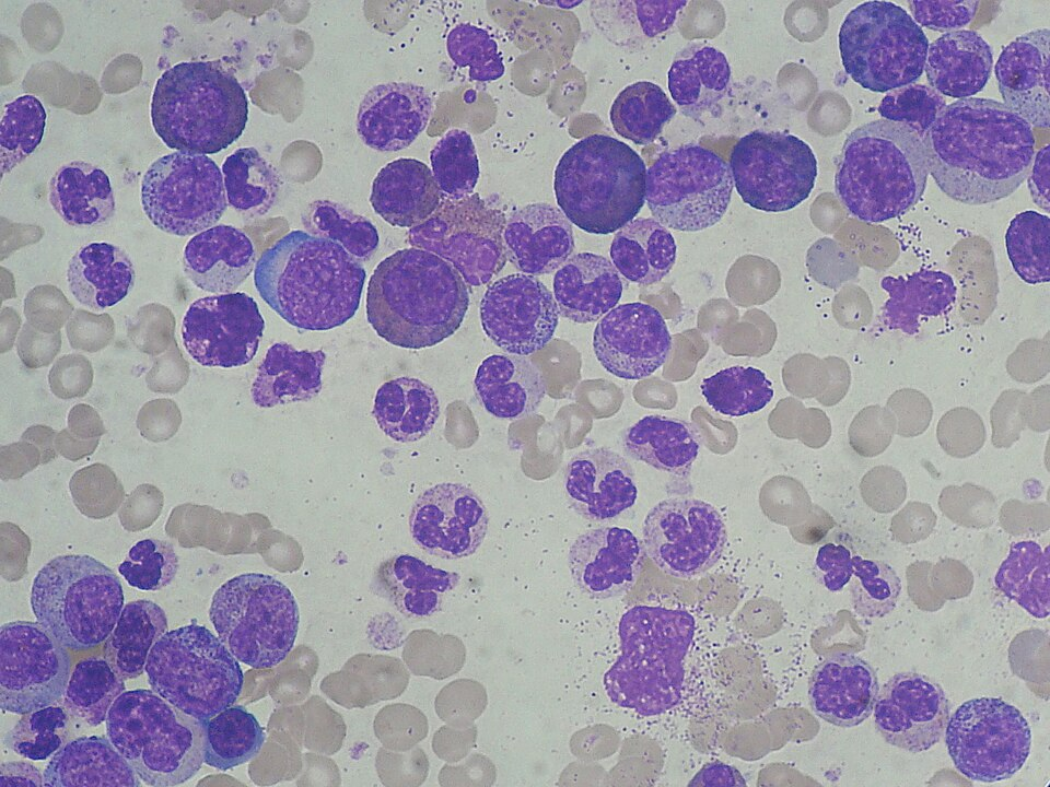
Chronic myeloid leukemia blood smear. Source: Paulo Henrique Orlandi Mourão, Wikimedia Commons. Licensed under CC BY-SA 3.0. https://commons.wikimedia.org/wiki/File:Chronic_Myeloid_Leukemia_smear_2009-04-09.JPG
Genetics: CML is associated with a specific genetic abnormality known as the Philadelphia chromosome, which forms when parts of two chromosomes swap places. This mutation causes uncontrolled cell growth in the bone marrow. Therefore, proof of diagnosis is attained by demonstration of the Philadelphia (Ph) chromosome (22q–) resulting from the balanced translocation t(9; 22) (q34;q11), and/or the BCR–ABL rearrangement in peripheral blood or bone marrow cells (Baccarani et al. 2009).
Vocabulary
References
Baccarani M, Dreyling M; ESMO Guidelines Working Group. Chronic myelogenous leukemia: ESMO clinical recommendations for diagnosis, treatment and follow-up. Ann Oncol. 2009 May;20 Suppl 4:105-7. doi: 10.1093/annonc/mdp143. PMID: 19454424.
Bryant BRE, Gardner JA, Devitt KA. Educational Case: Chronic Myeloid Leukemia. Acad Pathol. 2019 Dec 9;6:2374289519893084. doi: 10.1177/2374289519893084. PMID: 31840048; PMCID: PMC6902376.
Kimball, J.W. (n.d.). Chronic myelogenous leukemia (CML). LibreTexts Biology. https://bio.libretexts.org/Bookshelves/Introductory_and_General_Biology/Biology_(Kimball)/12%3A_Cancer/12.07%3A_Chronic_Myelogenous_Leukemia_(CML)
MedlinePlus. (n.d.). Complete blood count (CBC). U.S. National Library of Medicine. https://medlineplus.gov/lab-tests/complete-blood-count-cbc/
OpenStax. (n.d.). Leukocytes and platelets. In Anatomy and Physiology 2e. Rice University. https://openstax.org/books/anatomy-and-physiology-2e/pages/18-4-leukocytes-and-platelets
National Cancer Institute. (n.d.). Chronic myelogenous leukemia treatment (PDQ®)–Patient version. U.S. Department of Health and Human Services, National Institutes of Health. https://www.cancer.gov/types/leukemia/patient/cml-treatment-pdq